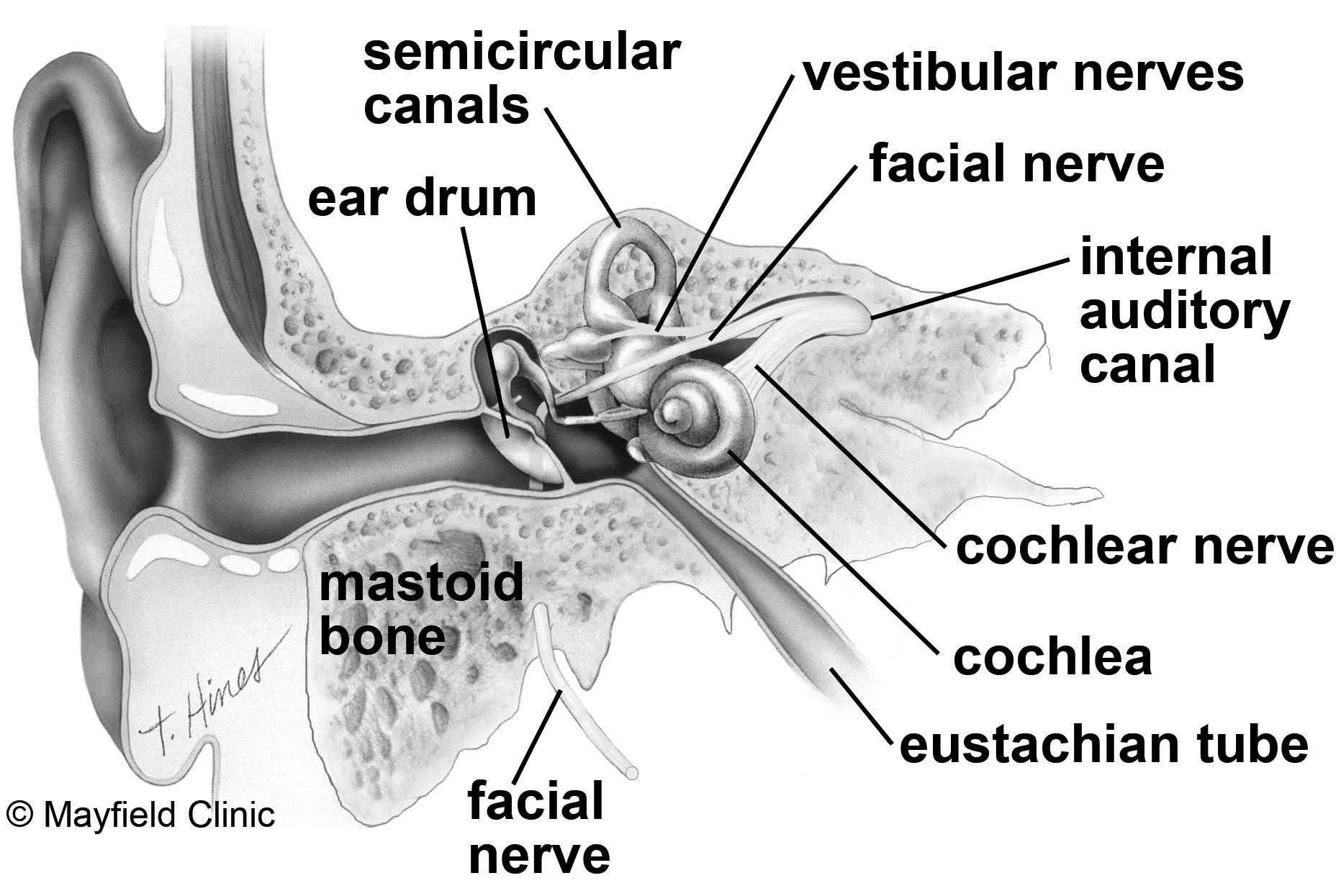
The symptoms caused by an acoustic neuroma follow the size and growth of the tumor. MRI scans of small (intracanalicular), medium, and large sizes of acoustic neuromas. Acoustic neuromas are classified according to their size as small (less than 1.5 cm), medium (1.5 to 2.5 cm), or large (more than 2.5 cm) (Fig. Eventually, the tumor can compress the brainstem. The pear-shaped tumor can continue to enlarge, compressing the trigeminal nerve, which is responsible for facial sensation. As the tumor grows, it expands from its origin inside the internal auditory canal out into the space between the brainstem and the bone known as the cerebellopontine angle. What is an acoustic neuroma?Īn acoustic neuroma, also called vestibular schwannoma, is a benign, slow-growing tumor that arises from the Schwann cells forming the sheath (covering) of the vestibulocochlear nerve. Eventually, the tumor compresses the brainstem. An acoustic neuroma expands out of the internal auditory canal, displacing the cochlear, facial, and trigeminal nerves. Likewise, facial sensation and feeling is controlled by the trigeminal nerve (fifth cranial nerve) and can be affected by large tumors.įigure 1. The close relationship of the vestibulocochlear and facial nerves explains why facial weakness can occur when an acoustic neuroma grows (Fig. The facial nerve (seventh cranial nerve) is responsible for moving the muscles of the face. Inside the canal, the vestibulocochlear nerve lies next to the facial nerve. The cochlear and vestibular nerves form a bundle inside the bony internal auditory canal before exiting to reach the brainstem. Electrical signals from the semicircular canals are carried to the brain by the superior and inferior vestibular nerves (responsible for balance). The three canals are able to sense head position and body posture. Attached to the cochlea are three semicircular canals positioned at right angles to each other. As the fluid moves, thousands of hair cells are stimulated, sending signals along the cochlear nerve, which are processed as hearing in the brain. The spiral-shaped cochlea is filled with liquid, which moves in response to vibrations. In turn, the stapes vibrates the oval window of the cochlea in the inner ear. The outer ear funnels sound down the ear canal to the eardrum, which vibrates three tiny bones called ossicles (malleus, incus, and stapes) in the middle ear (Fig. The vestibulocochlear nerve (eighth cranial nerve) is responsible for relaying hearing and balance signals from the inner ear to the brain. The middle and inner ear are located deep in the temporal bone of the skull. It consists of three parts: the outer ear, the middle ear, and the inner ear. The ear is our organ of hearing and balance.

Treatment options include observation, surgery, and radiosurgery. Because of their slow growth, not all acoustic neuromas need to be treated.


Over time the tumor can cause gradual hearing loss, ringing in the ear, and dizziness. Acoustic neuromas are benign (not cancer) and usually grow slowly. These tumors grow from the sheath covering the vestibulocochlear nerve. Acoustic neuroma (vestibular schwannoma) OverviewĪn acoustic neuroma is a tumor that grows from the nerves responsible for balance and hearing.


 0 kommentar(er)
